Even more complex and elegant than we thought

The language network is the brain’s “ambassador” 

Collectively responsible for articulating one's thoughts into verbal and written words, and comprehending and processing those of others, the language network serves one of the key cognitive functions that makes us human.

The language system has been described for decades. Neurosurgeons and neurologists often refer to it as an “eloquent region” of the brain; a key area worth protecting and examination to preserve healthy, independent function alongside vision and movement.

Yet, neurologically, language is not one functional process or region, but many. Language, after all, consists of multiple interdependent tasks. For example, when reading, listening, and speaking, visual, auditory, and motor processes are all required. Even these processes are just the bodily aspects of language. Additional processing is needed to comprehend the words on a page or the meaning of a sentence before articulating a coherent response. 

For this reason, it is not itself a “main network” of the brain as it is composed of parts of multiple other networks and therefore referred to at times as a “subnetwork”. This is also why increasing research into its crucial role reveals both more complexity and elegance than ever previously thought.

The future of language

Recent advances in neuroimaging and connectomics have vastly improved our understanding of language. Evaluating the functional and structural connectivity of the brain has aided in identifying close interactions and contributions from functional areas in the main brain networks that make up the language system.1

As such, using patient-specific brain maps, connectomics can pinpoint areas of specific function and help personalize operating and therapeutic decisions for individual patients.


Shifting to a network model of language

Neuroscience has long oscillated between a theory of equipotentiality - believing that the brain functions as a single faculty, and a theory of localization - suggesting that different traits or functions could be mapped onto specific areas of the brain.

In 1861, the discovery of “Broca’s area” — named for French surgeon Paul Broca — linked a specific cortical area in the left posterior inferior frontal gyrus with the function of word and speech production.2 Broca proposed the dedicated function of this area indicated the brain consisted of many organs with individual functions.3 German neurologist, Carl Wernicke, later expanded on Broca’s localizationist theory with his 1874 discovery of an area in the left posterior superior temporal gyrus that impacts language function. Later dubbed “Wernicke’s area,” this area is implicated in word-form recognition4 and phonological representation.5

The identification of these areas demonstrated that parts of the brain produced specific, eloquent function and should be avoided during surgery.6 Wernicke further proposed that since these two areas were both fundamentally involved in language processing, but were located in different regions of the brain, they must be interconnected.6

Nearly a century later, a German anatomist, Adolf Hopf, studied a small cortical area in the left dorsal premotor cortex7 known as 55b.8 He noted that significantly less myelin surrounded the neurons in this area,9 and it was consistently active during listening language tasks.9,10

Today, through the Human Connectome Project11 and research by Chang and others,12 neuroscience has shifted from Broca’s localizationist theory to a network model. Though there are localized, grey matter areas with specific functions, these areas are functionally connected to form highly correlated brain networks.13

These main networks each interact for prominent functions, like executive processing and motor capabilities. Yet, the network model also explains the correlation between areas from multiple networks working together to form integrated systems, like the language system.

A system of language functions

The main language functions activate a series of regions in the brain’s left hemisphere connected by white matter tracts. In addition to the arcuate fasciculus (AF) that connects Broca and Wernicke’s areas,14 white matter tracts connect areas from other networks.

The language system interacts with the visual system, auditory network, and sensorimotor network8 for reading, hearing, and producing speech. It also demonstrates functional connectivity to areas in the central executive network for specifying tasks, the dorsal attention network for maintained focus, and the default mode network for contemplation.

The language system consists of many discrete areas responsible for certain functions. As Broca and Wernicke’s areas indicate, areas can differentiate between the function of speech and the process of language.

Speech as a motor function causes the mouth and tongue to make comprehensive sounds in order to communicate. Language is an understanding of semantics and syntactic planning. To produce these cognitive processes, functional areas from different networks correlate with varying amounts of activity.

In a given moment, the language network activates functional areas in these networks to determine:

    • What sounds were heard
    • What words were read
    • The meaning of these sounds or words
    • What sounds should be made in response
    • The muscles needed to produce those sounds
    • The order of sounds required to produce words
    • How words should be organized to convey meaning through sentences

Each of the brain’s functional areas implicated in language processing fire together in different series and different amounts to comprehend and articulate language. To better understand where these areas and their white matter connections occur for individual patients, connectomic data can be used to model their interactions in a patient’s brain.

Language system location in the brain

Neuroscience’s understanding of language has greatly evolved over the past decade, and further refinement will continue to develop. Today, it is recognized that the functional areas of the brain implicated in language function occur in the lateral prefrontal cortex, left temporal regions beyond Wernicke's area, and the left dorso‐medial prefrontal cortex (dmPFC) for text comprehension.15 Areas in the inferior frontal gyrus, superior temporal cortex, and the anterior part of the left inferior temporal gyrus have also been implicated in language comprehension and production.16

While these areas are located in the left hemisphere, there is evidence for right hemisphere contributions.15 Right hemisphere activations have been noted while testing specific language processes, such as metaphor comprehension.15 This may indicate that right hemisphere contributions are task dependent, however there is still more to learn about bilateral impact in the language system.

It has also been proposed that specific areas are indispensable “hubs” for language production. Two potential language hubs may be areas STSvp and PHT.22 They are likely hub candidates due to their extensive functional connectivity to other brain regions, their direct connections to language production cortices, and their close geographic proximity to other language sites.22 While this hypothesis requires additional study,22 it does suggest that these areas be strongly considered and safeguarded during surgery.

LANGUAGE_AXI AXIAL VIEW

LANGUAGE_SAG SAGITTAL VIEW

LANGUAGE_COR CORONAL VIEW

Applying connectomics to language

Neurosurgery has long used awake brain surgery to gauge a patient’s ability to speak. However, while this method offers a chance to preserve speech function, it does not necessarily safeguard language function. Even if a patient maintains the ability to speak following a surgery, they may still encounter challenges with word association, object naming, or word choice.

To find data-driven methods of locating critical areas of speech and language function, functional magnetic resonance imaging (fMRI) can be used for noninvasive brain mapping to develop a connectomic atlas of the neural connections in a patient’s brain.6

Using connectomic data, neurosurgeons can evaluate areas of the brain — like Wernicke’s area and the surrounding grey matter areas — to determine which regions are responsible for language comprehension, semantic function, auditory recognition, and others. Connectomics provides a level of granularity previously unavailable to the field.

Armed with a connectomic atlas, neurosurgeons can analyze the functional areas and white matter tracts in a patient’s brain to determine where language function occurs.

These areas can then be compared and weighed against other important functional areas before planning the least disruptive path to a surgical site.

Why the connectome of language matters

A lesion or stroke can damage core cognitive ability, like language function, following injury to grey matter functional areas and/or the white matter tracts between them.

If you consider the brain’s connectome as a geographic map of the globe, each of the functional areas would be individual countries. The many highways connecting the countries would represent the brain’s white matter tracts. Now imagine one of the highways sustaining heavy damage and was no longer open for travel. The closure of this transportation route would have major effects on travel, shipping, trade, and other repercussions. It would not only affect the highway itself, but would have unforeseen ramifications for the connected countries.

The same is true for functional areas of the brain. Using connectomics, we can see not only which “highways” are important for transporting information, but also how they connect different “countries” and the possible cognitive effects that may result from damage to these areas.

Impacts from loss of language function

Damage to functional areas and white matter tracts13 of the language system can result in problems with language, speech, word association, recognition and familiarity,22 or naming.23 Injury to this system has also been implicated in neurological conditions and psychiatric disorders.

Stroke

In stroke patients, the location of the stroke plays a large factor on the severity of the damage. Since the language system is widespread in the left hemisphere, strokes in the lower left parts of the brain may cause greater amounts of language or speech dysfunction. Functional areas TE1a, TE1m, and TE1p are located in this region, and due to their implication in language processing tasks,24 can result in difficulty with language following a stroke.

Language disorders following a stroke often include alexia, agraphia, and aphasia.25 Alexia is a type of sensory aphasia where an individual loses the ability to read,25 whereas agraphia is the loss of writing skills.25 Difficulty writing may manifest from problems with language processing, visual perception, motor planning,25 or a combination of factors.

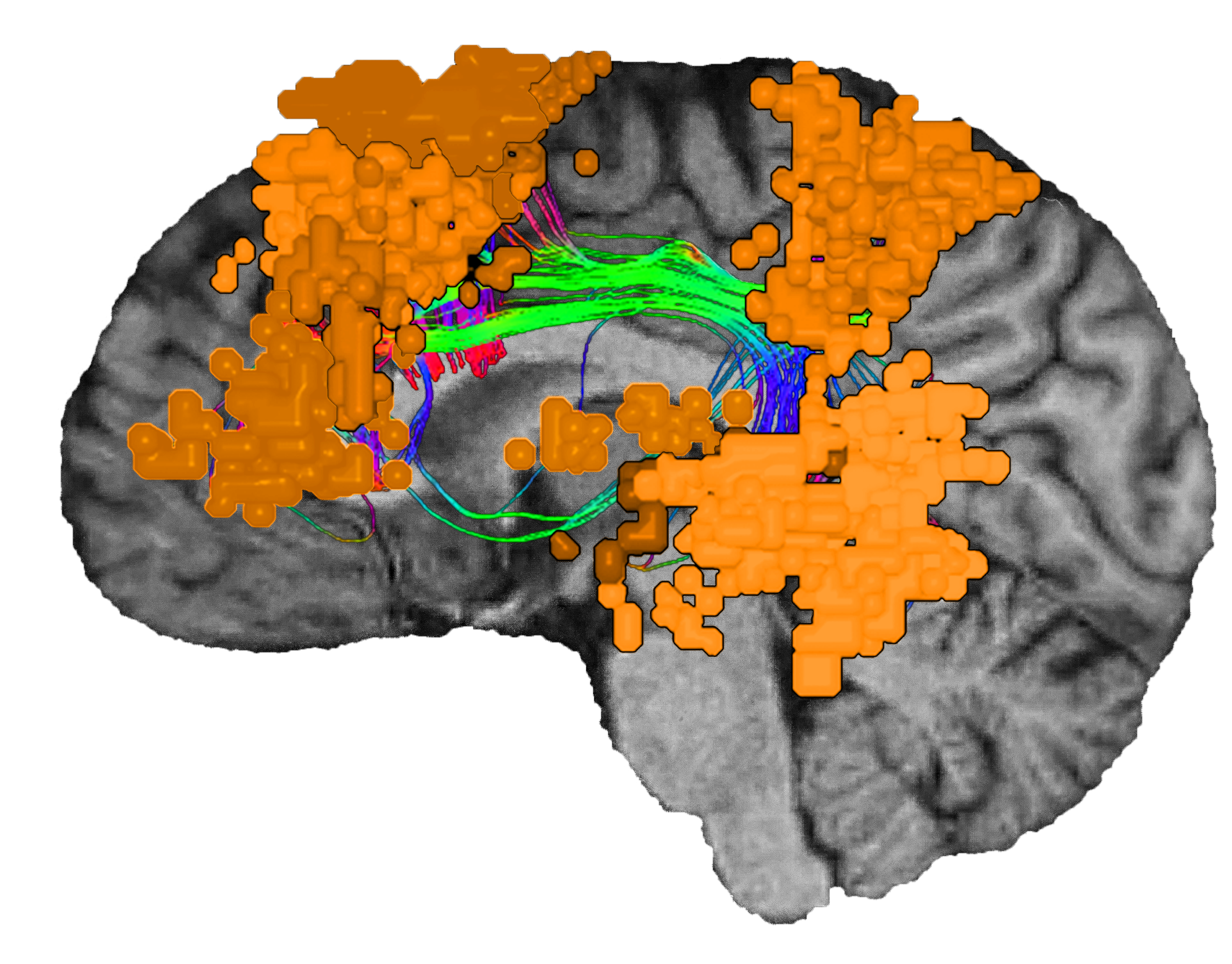
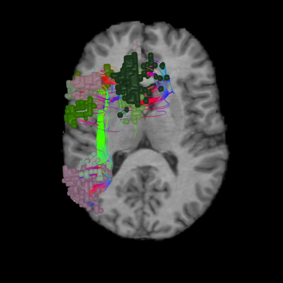
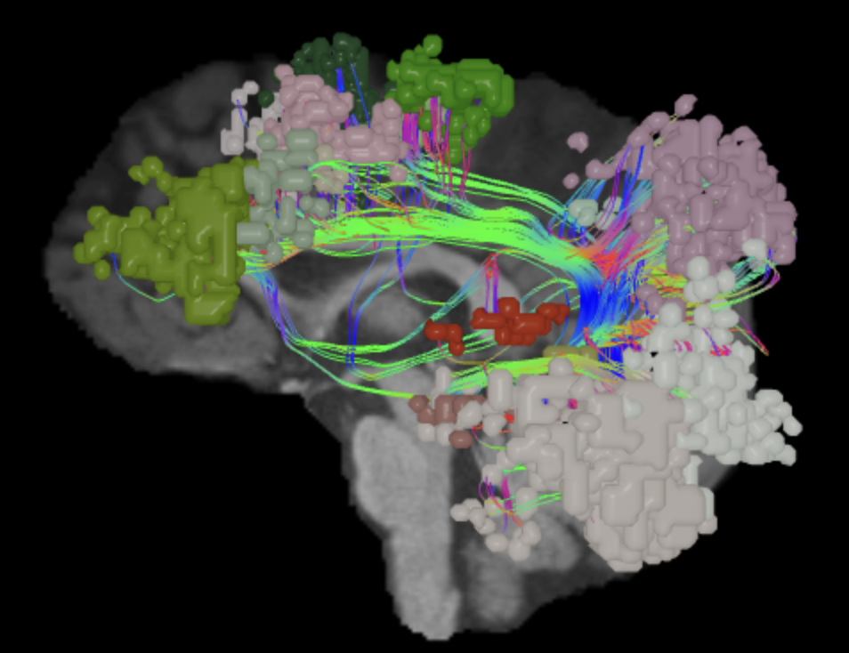
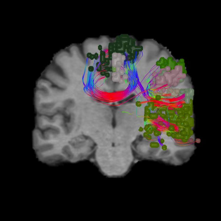
Aphasia is the loss or impairment of verbal communication,25 but it has been demonstrated to cause problems with writing and reading, as well as listening or speaking. Aphasia is most commonly caused by stroke, but may also result from a lesion in the left hemisphere25 or damage to the white matter tracts connecting regions of language in the brain — including the articulate fasciculus (AF), inferior longitudinal fasciculus (ILF), and the inferior fronto-occipital fasciculus (IFOF).6

However, in some cases, aphasia may also be a progressive, degenerative disease. Chawluk and colleagues26 have also suggested that progressive aphasia may occur without dementia and as a separate clinical syndrome from Alzheimer’s disease.26

Semantic dementia

Semantic dementia (SD) is the result of frontotemporal lobar degeneration27 typically occurring asymmetrically in the left hemisphere.28 Ventrorostal temporal lesions are also associated with the degeneration of white matter tracts connecting language areas29 that may lead to SD. Patients with semantic dementia have shown lower functional connectivity in the left uncinate fasciculus (UF), which has been previously linked to language functionality.30,31

SD is characterized by progressive aphasia — particularly anomia — and difficulty comprehending the meaning of words.27 Progressive stages of the disease have also shown atrophy near the contralateral temporal pole and medial prefrontal cortices which can result in social and behavioral changes.28

Unfortunately, early identification of SD can be difficult with standard clinical and neuropsychological evaluations.27 Using neuroimaging and task-related fMRI studies may offer opportunities to identify biomarkers of disease progression28 that may lead to earlier diagnoses and possible therapies.27

Epilepsy

Epilepsy has been shown to impact semantic memory in patients with varying outcomes based on whether epilepsy occurs in the left or right temporal lobe.32 Functional areas TGd and STGa play an important role in semantic memory,33,34,35 which will require further research on their possible implications in epilepsy.

Drane and colleagues36 found that patients experiencing right temporal lobe epilepsy (TLE) and post-surgical deficits demonstrate difficulty with face recognition and familiarity judgements.36 Since functional areas of the language system are primarily located in the left hemisphere, patients with left TLE unsurprisingly demonstrate difficulty with face naming.36 However, they did not exhibit problems with recognition or familiarity.36

Performing surgical treatment for TLE may reduce seizures, but it relies on accurate lateralization of the TLE. Resting-state fMRI has been useful for mapping connectivity and determining left or right lateralized TLE. Morgan and colleagues identified an area in the ventral lateral nucleus of the right thalamus with functional connectivity to the hippocampi that may be an indicator of lateralization and distinguish left from right TLE subjects.37 Predicting hemispheric language dominance helps surgeons preserve the greatest amount of language function following an operation.

Connectomics offers the opportunity to define key functional language areas and their white matter connections with great specificity. In addition to understanding where primary areas of language reside in each patient’s brain, continued research may also lead to more specific models of semantic memory and the localization of seizures to improve post-surgical outcomes.32

Since functional areas and white matter tracts of the language system make up a highly interconnected system, developing patient-specific connectomic atlases will aid in diagnosing patients13 and providing greater opportunity for therapy or treatment.

Emerging concepts of language

While it is widely accepted that language is a primarily left lateralized function, newer concepts of language function have been slowly evolving. Research has pointed to a possible dual-stream model38 of language (similar to the visual system) consisting of a dorsal and ventral stream39,40 within the language system.6

This idea has been further refined to suggest that there may be two dorsal and two ventral pathways41 connecting the prefrontal and temporal areas of the language system. The dorsal streams connect the temporal and premotor cortices, as well as the temporal cortex and Broca’s area.41 The ventral streams follow the UF and the IFOF.41

Further fMRI and tractography studies will continue to shed light on the flow of information through these white matter tracts and their relationships with language-focused functional areas.6,42 In addition to studying well-known language regions, research has also begun questioning whether language is solely localized in the left hemisphere. Some white matter tracts show bilateral connections, and Thiel and colleagues43 have found that patients with left-lateralized, slow-growing lesions may have opportunity to develop language function in areas of the right hemisphere homologous to left brain language areas.43

Since the process of language remains a complex and multifaceted function, there is still much research to be done in this area. Yet, with the added insight from connectomic data, neurosurgeons may begin incorporating cutting-edge research for patient support right away.

The language system is just one piece of the complex networks that make up the human brain. Discover the key roles of other brain networks and how they interconnect with the language system.

Reference list

Expand for full list of references
  1. Fedorenko E, Thompson-Schill S. Reworking the language network. Trends in Cognitive Science. 2014;18(3):120-126. doi: 10.1016/j.tics.2013.12.006.
  1. Flinker A, Korzeniewska A, Shestyuk A, Franaszczuk P, Dronkers N, Knight R, Crone N. Redefining the Broca’s area in speech. Proc Natl Acad Sci U S A. 2015;112(9): 2871–2875. doi:10.1073/pnas.1414491112.
  1. Keller SS, Crow T, Foundas A, Amunts K, Roberts N. Broca's area: nomenclature, anatomy, typology and asymmetry. Brain Lang. 2009;109(1):29-48. doi:10.1016/j.bandl.2008.11.005.
  1. DeWitt I, Rauschecker J. Wernicke’s area revisited: Parallel streams and word processing. Brain Lang. 2013;127(2):181-191. doi:10.1016/j.bandl.2013.09.014.
  1. Linell P. The concept of phonological form and the activities of speech production and speech perception. J Phon. 1982;10(1):37-72. doi:10.1016/S0095-4470(19)30910-6
  1. Monroy-Sosa A, Chakravarthi S, Cortes-Contreras A, et al. The Evolution of Cerebral Language Localization: Historical Analysis and Current Trends. World Neurosurg. 2021;145:89-97. doi.org/10.1016/j.wneu.2020.09.028.
  1. Chang EF, Kurteff G, Andrews JP, et al. Pure apraxia of speech after resection based in the posterior middle frontal gyrus. Neurosurg. 2020;87(3):E383–E389. doi.org/10.1093/neuros/nyaa002.
  1. Briggs RG, Conner AK, Baker CM, et al. A Connectomic Atlas of the Human Cerebrum-Chapter 18: The Connectional Anatomy of Human Brain Networks. Oper Neurosurg (Hagerstown). 2018;15(suppl_1):S470-S480. doi:10.1093/ons/opy272.
  1. Brodwin E. This mind-blowing brain map accomplishes something that scientists have been working on for 100 years. Business Insider. https://www.businessinsider.com.au/new-brain-map-accomplishes-what-scientists-have-been-working-on-for-100-years-2016-7?r=US&IR=T. Accessed April 29, 2021.
  1. Glasser M, Coalson T, Robinson E, et al. A multi-modal parcellation of human cerebral cortex. Nature 536. 2016. https://doi.org/10.1038/nature18933.
  1. Human Connectome Project. Mapping the human brain connectivity. www.humanconnectomeproject.org. Accessed October 16, 2020.
  1. Chang EF, Raygor KP, Berger MS. Contemporary model of language organization: an overview for neurosurgeons. Journal of Neurosurgery JNS. 2015;122(2):250-261. doi.org/10.3171/2014.10.JNS132647.
  1. Bajada CJ, Ralph MAL, Cloutman LL. Transport for language south of the Sylvian fissure: The routes and history of the main tracts and stations in the ventral language network. Cortex. 2015;69;141-151. doi.org/10.1016/j.cortex.2015.05.011.
  1. Catani M, Allin MPG, Husain M, et al. Symmetries in human brain language pathways correlate with verbal recall. Proc Natl Acad Sci U S A. 2007;104(43):17163-17168. doi: 10.1073/pnas.0702116104.
  1. Ferstl EC, Neumann J, Bogler C, von Cramon DY. The extended language network: a meta-analysis of neuroimaging studies on text comprehension. Hum Brain Mapp. 2008;29(5):581-593. doi:10.1002/hbm.20422.
  1. Papathanassiou D, Etard O, Mellet E, Zago L, Mazoyer B, Tzourio-Mazoyer N. A Common Language Network for Comprehension and Production: A Contribution to the Definition of Language Epicenters with PET. NeuroImage. 2000;11(4);347-357. doi.org/10.1006/nimg.2000.0546.
  1. Hagoort P. On Broca, brain, and binding: a new framework. Trends Cogn Sci. 2005;9(9):416-423. doi:10.1016/j.tics.2005.07.004.
  1. Venezia JH, Fillmore P, Matchin W, Isenberg AL, Hickok G, Fridriksson J. Perception drives production across sensory modalities: A network for sensorimotor integration of visual speech. Neuroimage. 2016;126:196-207. doi:10.1016/j.neuroimage.2015.11.038.
  1. Baker CM, Burks JD, Briggs RG, et al. A Connectomic Atlas of the Human Cerebrum—Chapter 3: The Motor, Premotor, and Sensory Cortices. Operative Neurosurgery. 2018;15(suppl_1):S75–S121. doi.org/10.1093/ons/opy256.
  1. Gabrieli JDE. Cognitive neuroscience of human memory. Annu Rev Psychol. 1998;49(1):87-115. doi.org/10.1146/annurev.psych.49.1.87.
  1. Pammer K, Hansen P, Holliday I, Cornelissen P. Attentional shifting and the role of the dorsal pathway in visual word recognition. Neuropsychologia. 2006;44(14):2926-36. doi:10.1016/j.neuropsychologia.2006.06.028.
  1. Baker CM, Burks JD, Briggs RG. et al. A Connectomic Atlas of the Human Cerebrum—Chapter 6: The Temporal Lobe. Operative Neurosurgery. 2018;15(suppl_1):S245–S294. doi.org/10.1093/ons/opy260.
  1. Eichenbaum H, Yonelinas AP, Ranganath C. The medial temporal lobe and recognition memory. Annu Rev Neurosci. 2007;30:123-152. doi:10.1146/annurev.neuro.30.051606.094328.
  1. Glosser G, Salvucci AE, Chiaravalloti ND. Naming and recognizing famous faces in temporal lobe epilepsy. Neurology. 2003;61(1):81-86. doi:10.1212/01.WNL.0000073621.18013.E1.
  1. Glasser MF, Coalson TS, Robinson EC et al. A multi-modal parcellation of human cerebral cortex. Brain. 2016;536(7615):2093-2107.
  1. Sinanović O, Mrkonjić Z, Zukić S, Vidović M, Imamović K. Post-stroke language disorders. Acta Clin Croat. 2011;50(1):79-94.
  1. Chawluk JB, Mesulam MM, Hurtig H, et al. Slowly progressive aphasia without generalized dementia: studies with positron emission tomography. Ann Neurol. 1986;19(1):68-74. doi:10.1002/ana.410190112.
  1. Czarnecki K, Duffy JR, Nehl CR, et al. Very early semantic dementia with progressive temporal lobe atrophy: an 8-year longitudinal study. Arch Neurol. 2008;65(12):1659-1663. doi:10.1001/archneurol.2008.507.
  1. Landin-Romero R, Tan R, Hodges JR, Kumfor F. An update on semantic dementia: genetics, imaging, and pathology. Alzheimers Res Ther. 2016;8(1):52. doi:10.1186/s13195-016-0219-5.
  1. Acosta-Cabronero J, Patterson K, Fryer TD, et al. Atrophy, hypometabolism and white matter abnormalities in semantic dementia tell a coherent story. Brain. 2011;134(7):2025–2035. doi.org/10.1093/brain/awr119.
  1. Agosta F, Galantucci S, Canu E et al. Disruption of structural connectivity along the dorsal and ventral language pathways in patients with nonfluent and semantic variant primary progressive aphasia: A DT MRI study and a literature review. Brain Lang. 2013;127(2):157-166. doi: 10.1016/j.bandl.2013.06.003.
  1. Agosta F, Henry RG, Migliaccio R et al. Language networks in semantic dementia. Brain. 2010;133(1):286-299. doi: 10.1093/brain/awp233.
  1. Drane DL, Ojemann GA, Aylward E, et al. Category-specific naming and recognition deficits in temporal lobe epilepsy surgical patients. Neuropsychologia. 2008;46(5):1242-1255. doi:10.1016/j.neuropsychologia.2007.11.034.
  1. Olson IR, Von Der Heide RJ, Alm KH, Vyas G. Development of the uncinate fasciculus: Implications for theory and developmental disorders. Dev Cogn Neurosci. 2015;14:50-61. doi: 10.1016/j.dcn.2015.06.003.
  1. Catani M, Mesulam M. The arcuate fasciculus and the disconnection theme in language and aphasia: History and current state. Cortex. 2008;44(8):953-961. doi: 10.1016/j.cortex.2008.04.002.
  1. Grossman M, McMillan C, Moore P, et al. What’s in a name: voxel-based morphometric analyses of MRI and naming difficulty in Alzheimer’s disease, frontotemporal dementia and corticobasal degeneration. Brain. 2004;127(Pt3):628-649. doi: 10.1093/brain/awh075.
  1. Drane DL, Ojemann JG, Phatak V, et al. Famous face identification in temporal lobe epilepsy: support for a multimodal integration model of semantic memory. Cortex. 2013;49(6):1648-1667. doi:10.1016/j.cortex.2012.08.009.
  1. Morgan VL, Sonmezturk HH, Gore JC,Abou‐Khalil B. Lateralization of temporal lobe epilepsy using resting functional magnetic resonance imaging connectivity of hippocampal networks. Epilepsia. 2012;53:1628-1635. doi.org/10.1111/j.1528-1167.2012.03590.x.
  1. Hickok G, Poeppel D. Towards a functional neuroanatomy of speech perceptionTrends Cogn Sci. 2000;4:131-138.
  1. Parker GJ, Luzzi S, Alexander DC, Wheeler-Kingshott CA, Ciccarelli O, Lambon Ralph MA. Lateralization of ventral and dorsal auditory-language pathways in the human brain. Neuroimage. 2005;25:656-666.
  1. Duffau H, Gatignol P, Mandonnet E, Peruzzi P, Tzourio-Mazoyer N, Capelle L. New insights into the anatomo-functional connectivity of the semantic system: a study using cortico-subcortical electrostimulations. Brain. 2005;128:797-810.
  1. Friederici AD, Gierhan SME. The language network. Curr Opin Neurobiol. 2013;23(2):250-254. doi.org/10.1016/j.conb.2012.10.002.
  1. G. Wysiadecki, E. Clarke, M. Polguj, R. Haladaj, A. Zytkowski, M. Topol Klingler's method of brain dissection: review of the technique including its usefulness in practical neuroanatomy teaching, neurosurgery and neuroimaging. Folia Morphol (Warsz). 2019;78.455-466
  2. Thiel A, Habedank B, Herholz B, et al. From the left to the right: How the brain compensates progressive loss of language function. Brain Lang. 2006;98(1):57-65. doi.org/10.1016/j.bandl.2006.01.007