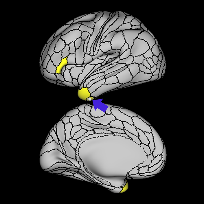
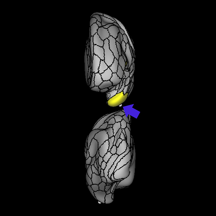
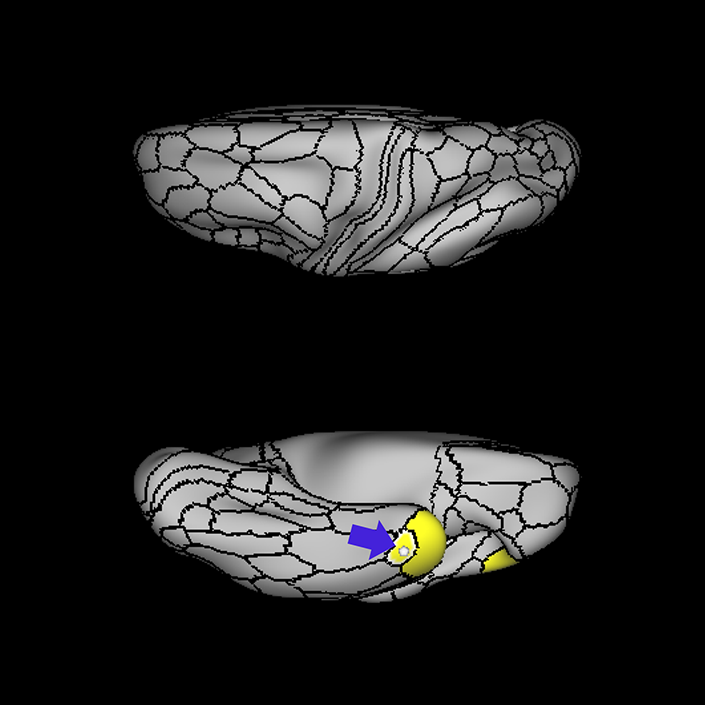
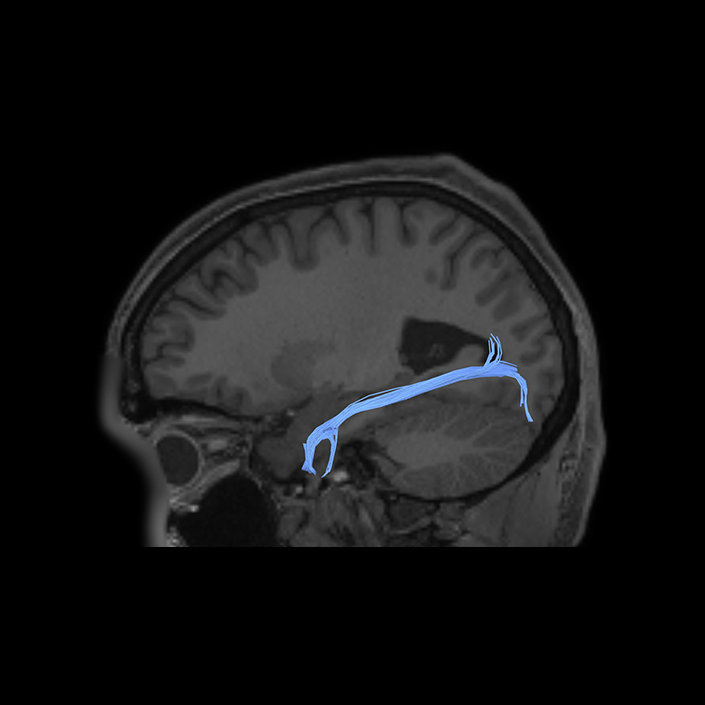
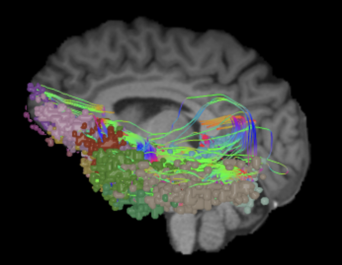
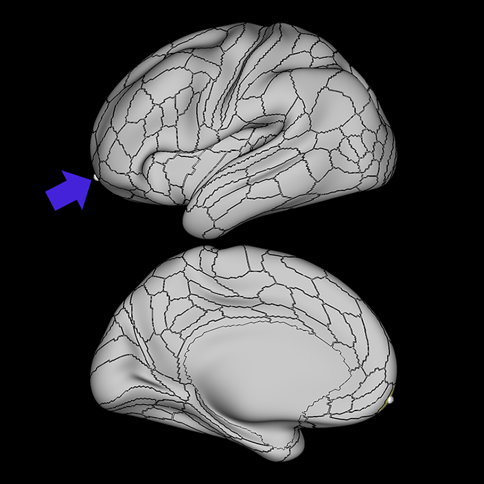
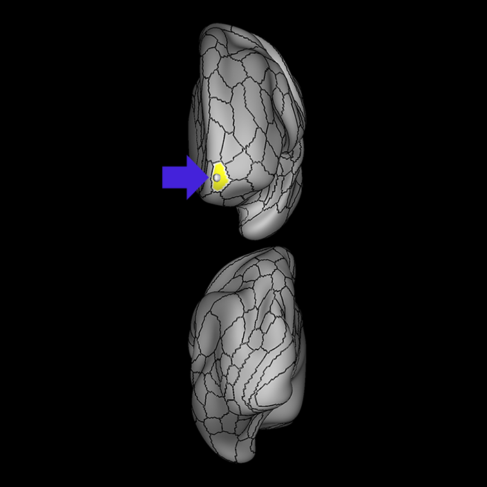
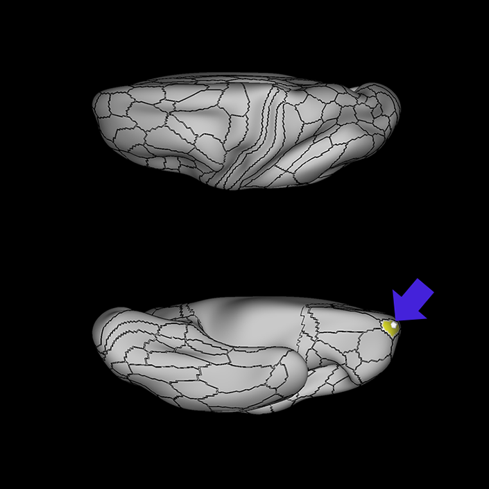
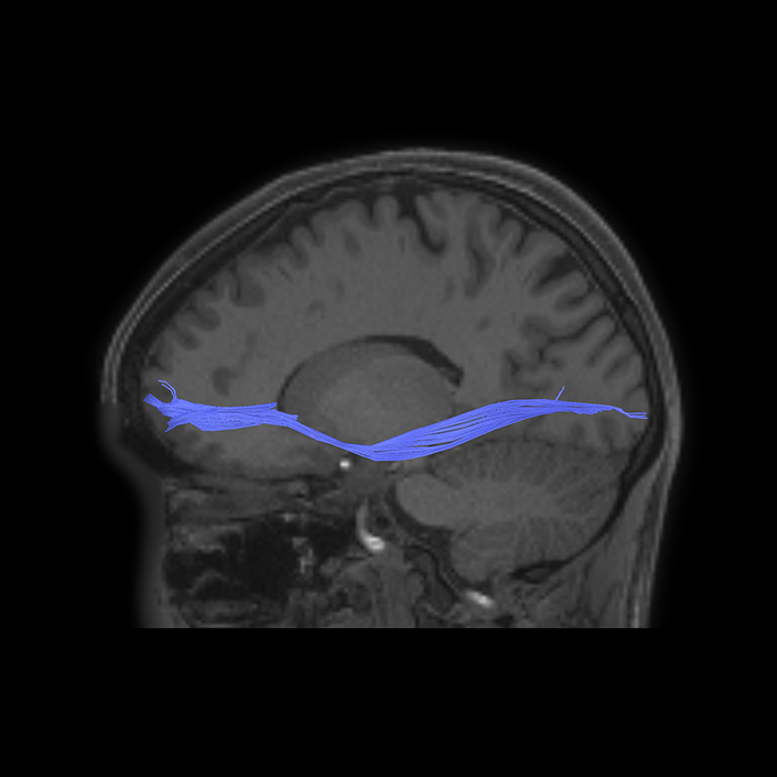
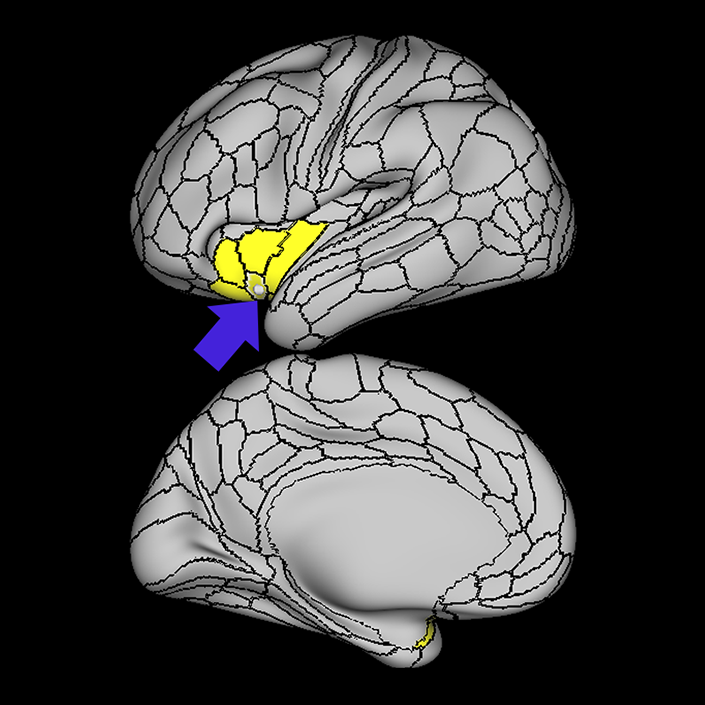
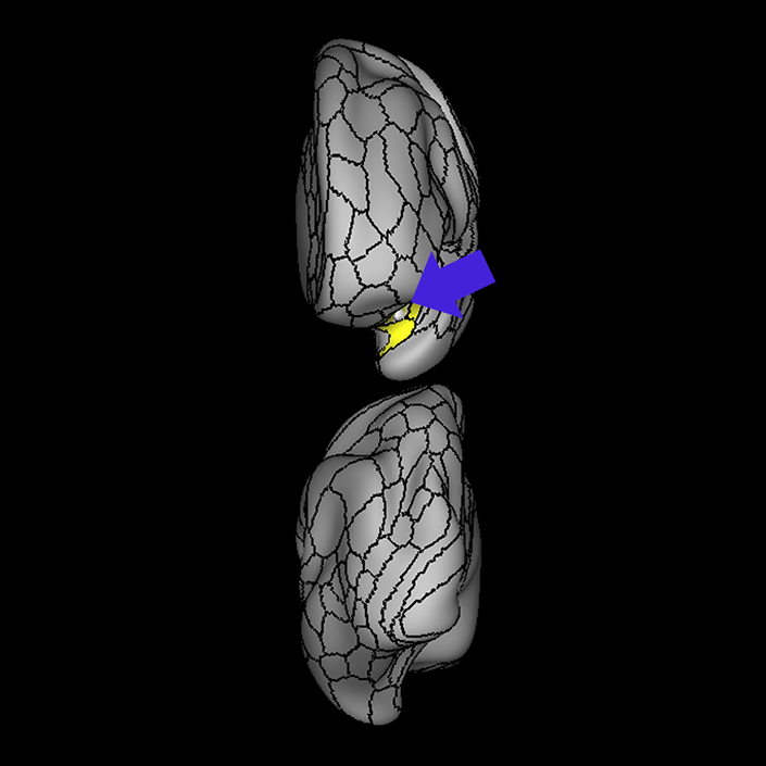
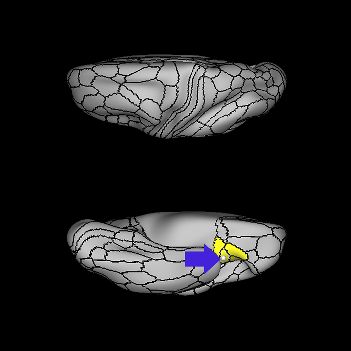
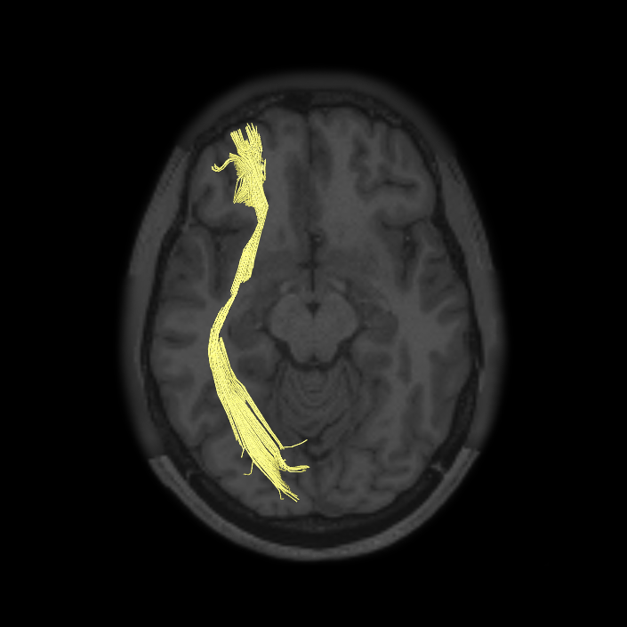
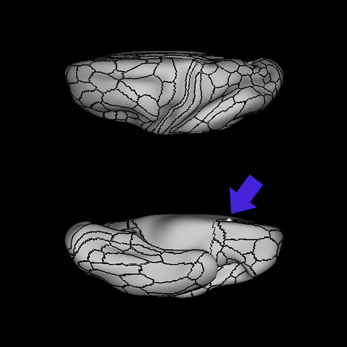
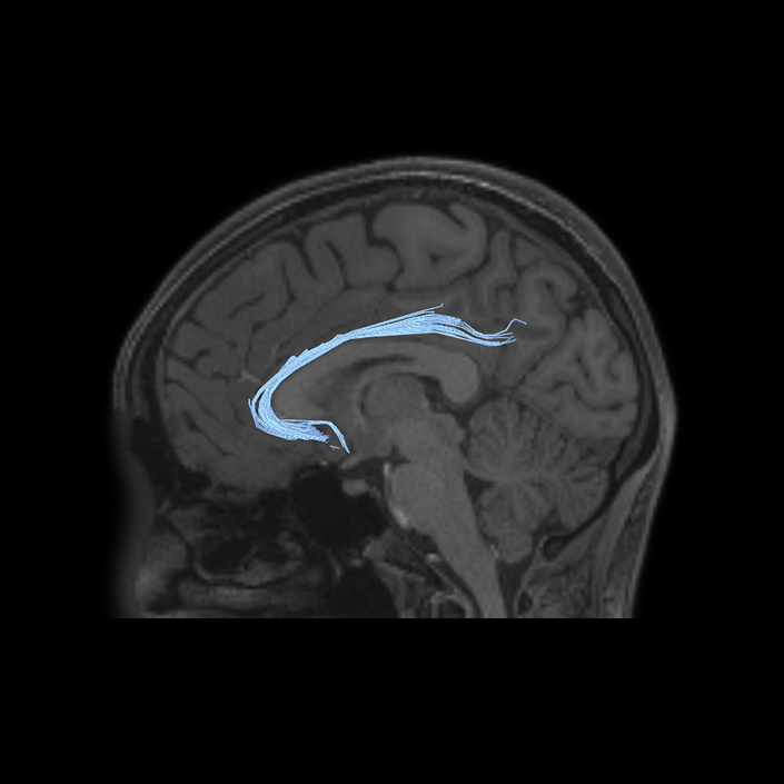
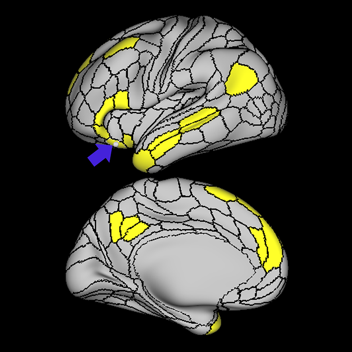
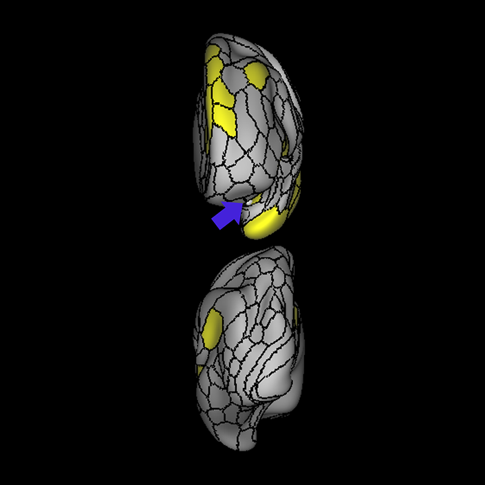
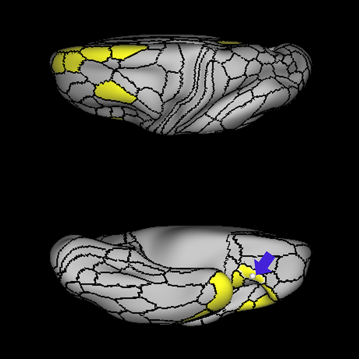
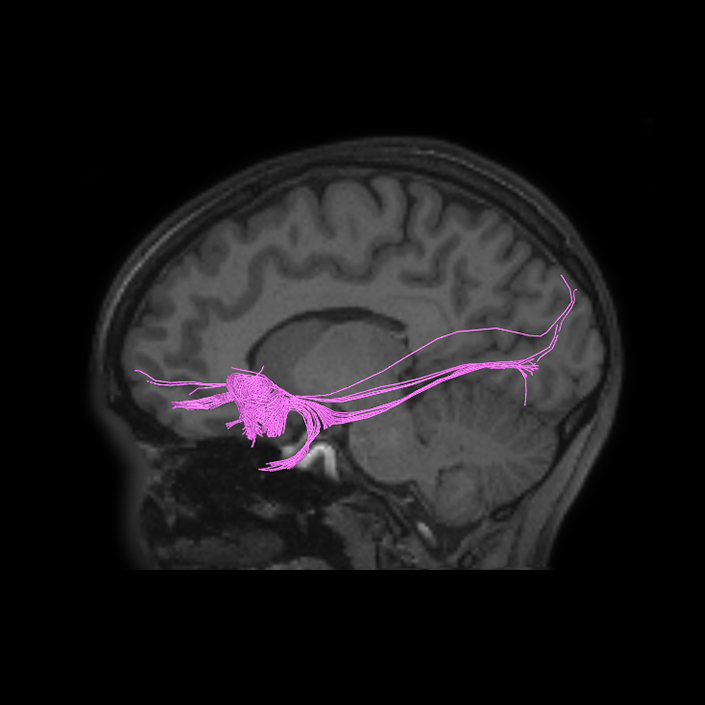
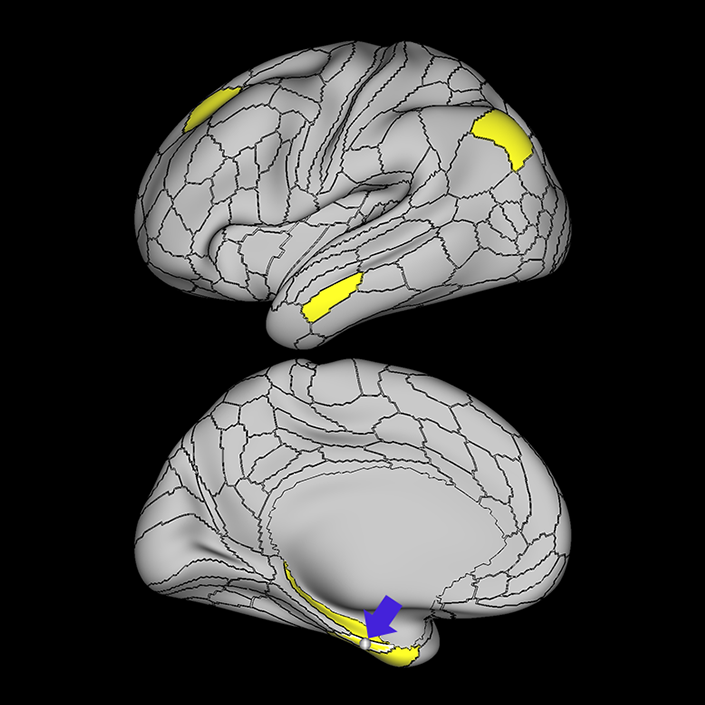
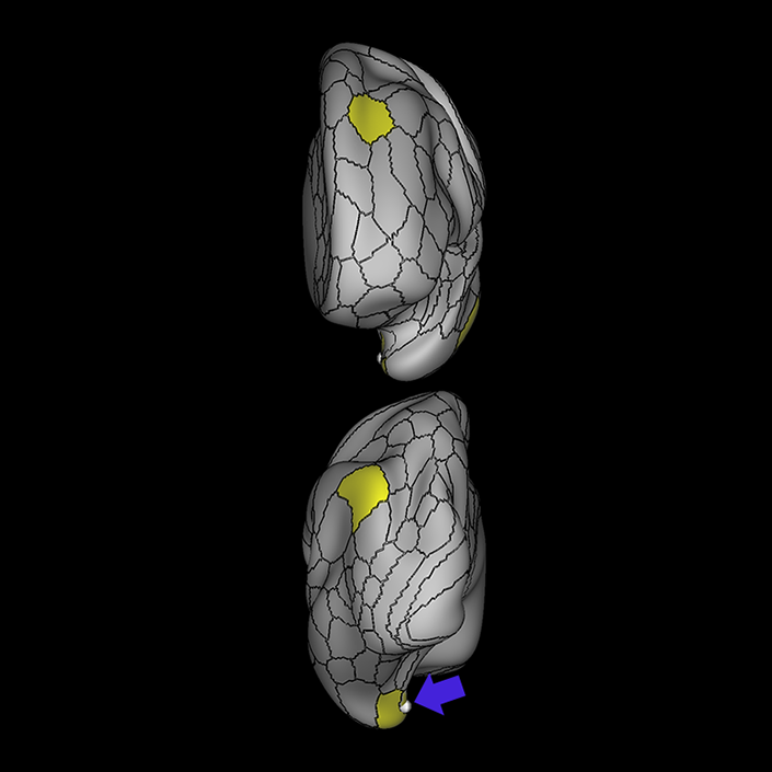
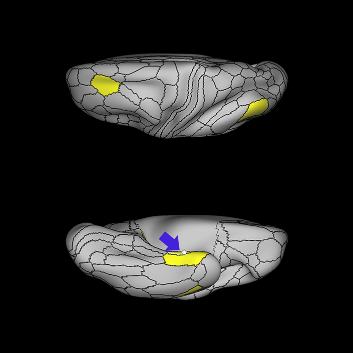
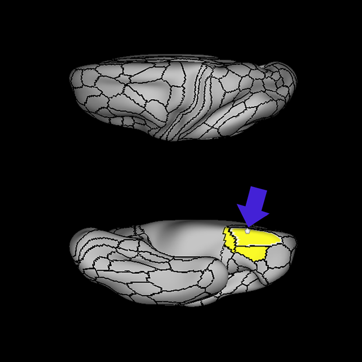
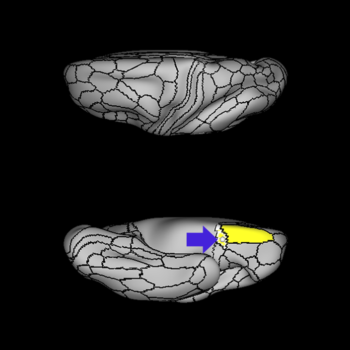
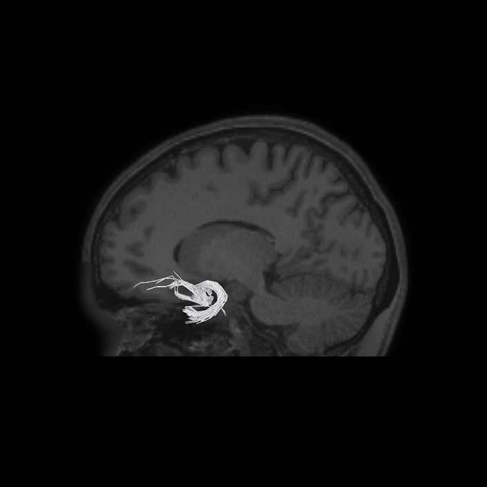
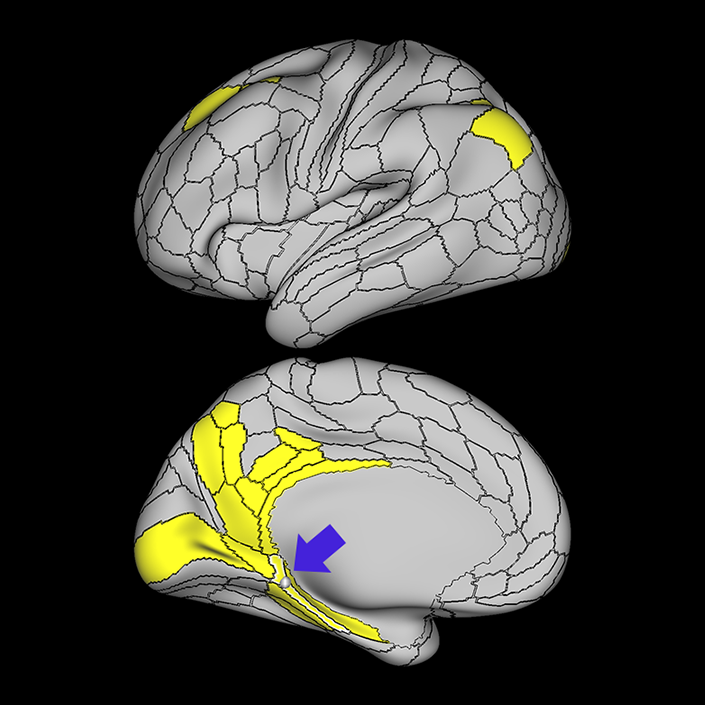
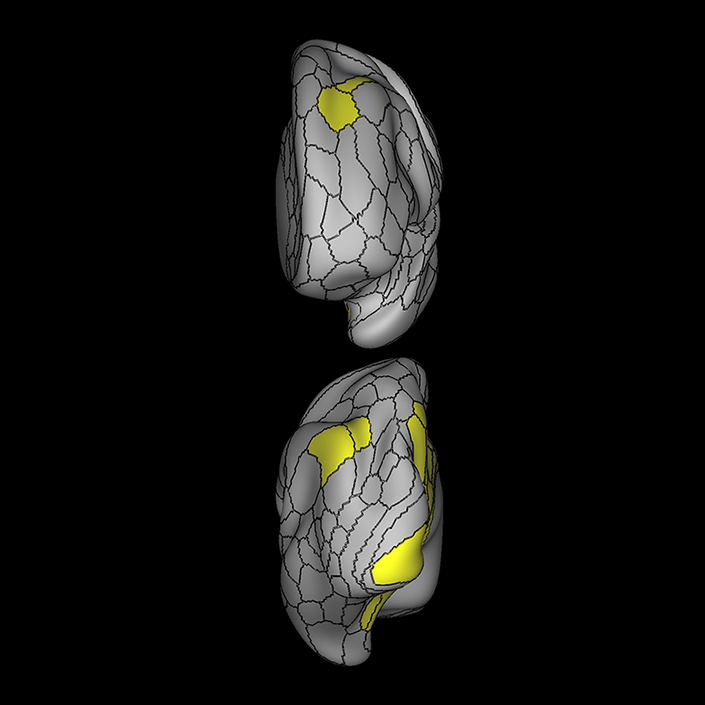
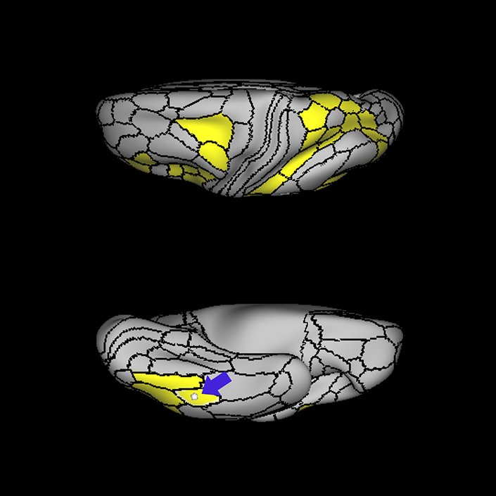
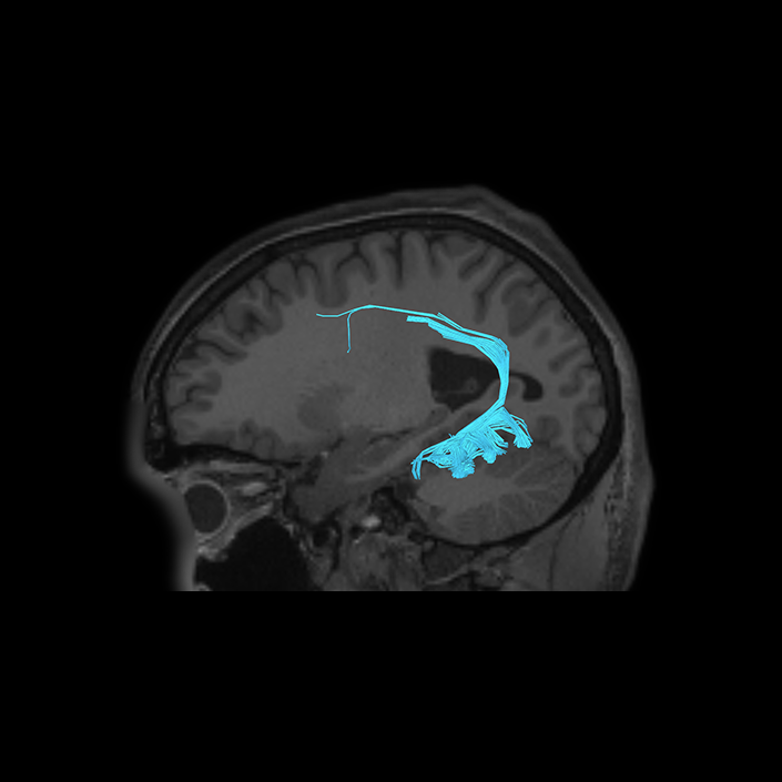
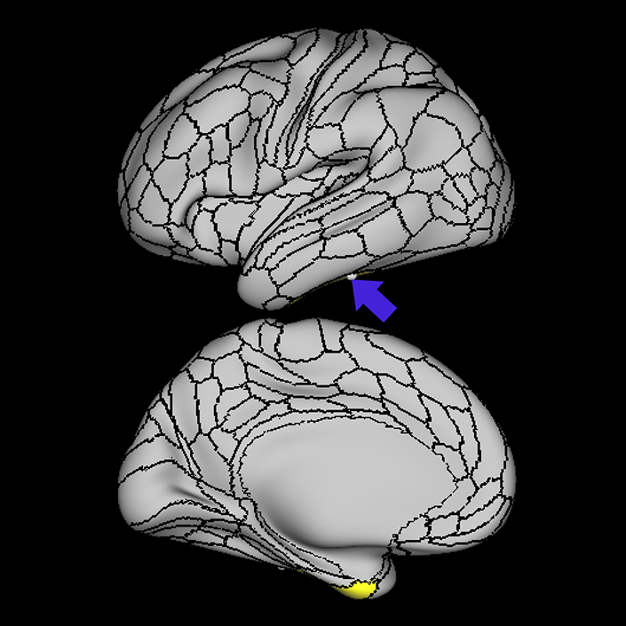
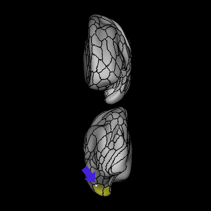
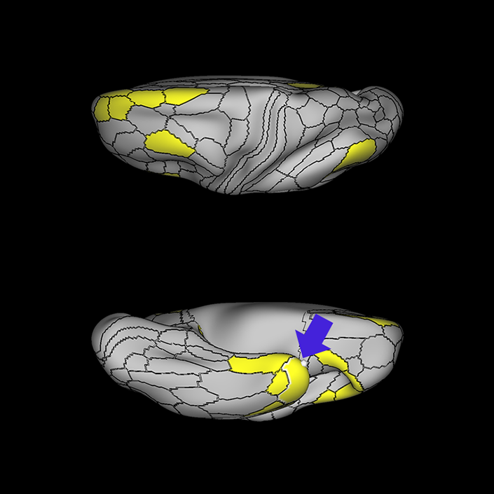
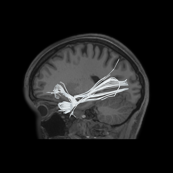
ᐅ SummaryArea 10pp (10 polar polar): part of frontal polar region of lateral temporal lobe. Involved in episodic and working memory tasks. Brodmann area 10 more generally is activated in relation to increasing complexity of working memory tasks. This area also plays a role in abstract cognitive function. ᐅ Where is it?Area 10pp is located at the anterior tip of the frontal pole. It is located at the fusiform junction of the anterior most aspects of the superior and middle frontal gyri. ᐅ What are its borders?Area 10pp borders the orbitofrontal region on its inferoposterior boundary, and area 10d on its superior boundary. Area a10p is its lateral neighbor. ᐅ What are its functional connections?Area 10pp lacks strong evidence of functional connectivity to other areas, at least when using a z-score cutoff of 20. There is borderline evidence of functional connectivity with its neighbor a10p, as well as areas 8Av and 8C in the posterior dorsolateral frontal lobe, areas 31pd and 31pv in the posterior cingulate cortex, areas PFm, and PGi in the inferior parietal lobule, and area STSvp in the superior temporal sulcus. ᐅ What are its white matter connections?Area 10pp is structurally connected to the IFOF and contralateral hemisphere. IFOF connections travel from 10pp through the extreme/external capsule and continue posteriorly to end at occipital lobe parcellations V1 and V2. Contralateral connections course through the genu of the corpus callousum with the forceps minor to terminate at 10d and 10pp. Local short association bundles are connected with 10d. ᐅ What is known about its function?Area 10pp is involved in episodic and working memory tasks. Brodmann area 10 more generally is activated in relation to increasing complexity of working memory tasks. This area also plays a role in abstract cognitive function. |
|
A: lateral-medial
B: anterior-posterior
C: superior-inferior
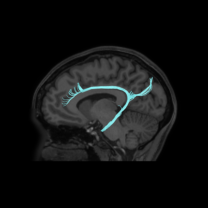
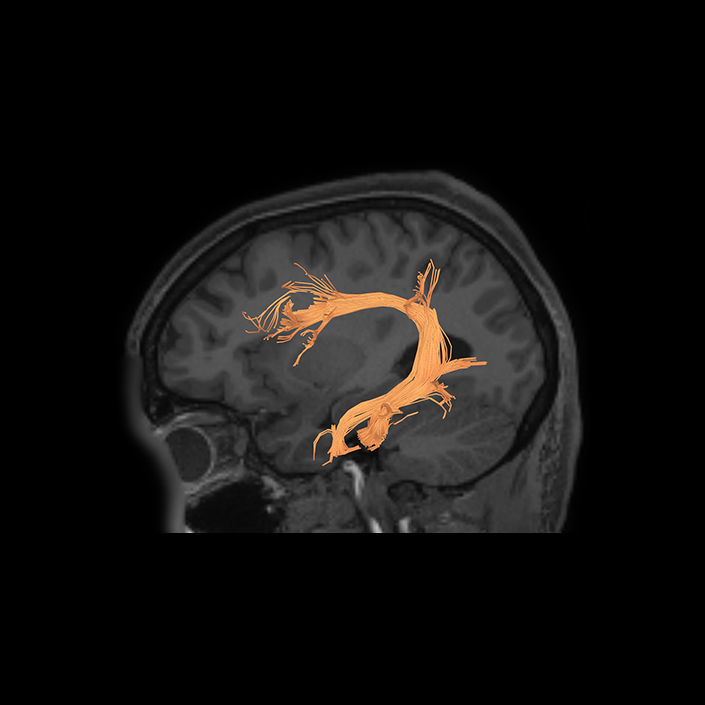
DTI image |
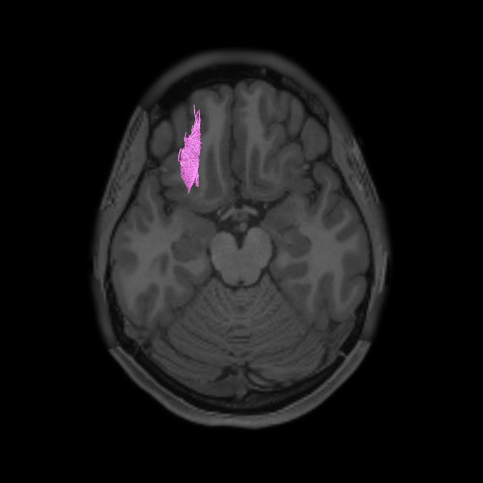
ᐅ SummaryArea 11: part of the the orbitofrontal regions. Research indicates its primary function is to receive, integrate, and modulate olfactory information to evaluate food-related reinforcers and analyze satiety and anticipation of food rewards. ᐅ Where is it?Area 11l is located in the anterior orbitofrontal cortex. ᐅ What are its borders?Area 11l borders area a10p anteriorly, area a47r laterally, areas 13l and 47m posteriorly, and OFC medially. ᐅ What are its functional connections?Area 11l demonstrates functional connectivity to areas 46, a9-46v, p9-46v and IFSa in the frontal lobe and IP2 in the parietal lobe. ᐅ What are its white matter connections?Area 11l is structurally connected to the occipital lobe through the inferior fronto-occipital fasciculus. In some individuals 11l also connects with the uncinate fasciculus but this tract is inconsistent. Inferior fronto-occipital fasciculus fibers project through the extreme/external to end at occipital lobe area V1. ᐅ What is known about its function?Research on area 11l indicates its primary function is to receive, integrate, and modulate olfactory information to evaluate food-related reinforcers and analyze satiety and anticipation of food rewards. |
|
A: lateral-medial
B: anterior-posterior
C: superior-inferior
DTI image |
ᐅ SummaryArea 13l: part of the the orbitofrontal regions. Has been implicated as a secondary hub for olfactory, gustatory, visceral and food texture processing, integrating this information to assess satiety based on a comparison between food reward and current internal states. ᐅ Where is it?Area 13l is located in the posterior orbitofrontal cortex. ᐅ What are its borders?Area 13l borders area 11l anteriorly, areas 47s and 47m posteriorly and laterally, and OFC medially. ᐅ What are its functional connections?Area 13l demonstrates functional connectivity to areas 47m and OFC. ᐅ What are its white matter connections?Area 13l is structurally connected to local parcellations. Short association bundles connect with 11l, 47m and 47s. ᐅ What is known about its function?Area 13l has been implicated as a secondary hub for olfactory, gustatory, visceral and food texture processing, integrating this information to assess satiety based on a comparison between food reward and current internal states. |
|
A: lateral-medial
B: anterior-posterior
C: superior-inferior
DTI image |


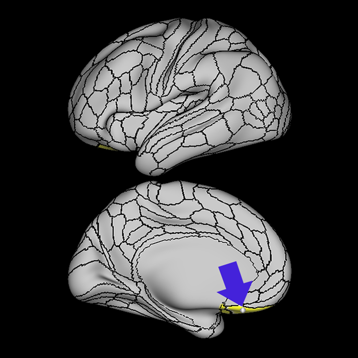
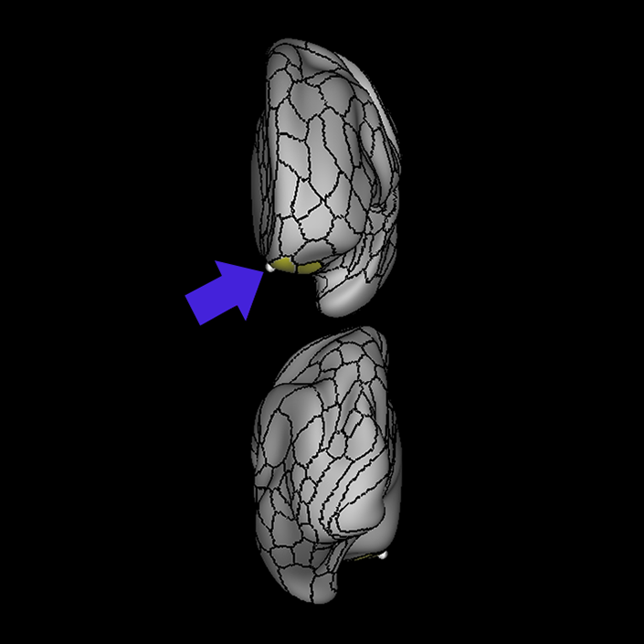
ᐅ SummaryArea 25: part of anterior cingulate regions. Has been implicated as part of the affect division" of the anterior cingulate cortex and has been linked to conditioned emotional learning ᐅ Where is it?Area 25 is located in the most posterior portion of the subcallosal area. ᐅ What are its borders?Area 25 borders area a24 and s32 anteriorly. Its inferior and posterior borders include the OFC and pOFC areas. ᐅ What are its functional connections?Area 25 demonstrates functional connectivity to area s32. ᐅ What are its white matter connections?Area 25 is connected to the cingulum. White matter tracts from this parcellation are variable. In some individulas tracts connect to anterior parcellations however, this is inconsistent across individuals. Fibers from 25 project posterior above the corpus callosum to v23ab and 31pv. ᐅ What is known about its function?Area 25 has been implicated as part of the "affect division" of the anterior cingulate cortex and has been linked to conditioned emotional learning, emotional expression, assessment of motivational content, assignment of emotional valence to internal and external stimuli, and maternal-infant interactions. |
|
A: lateral-medial
B: anterior-posterior
C: superior-inferior
DTI image |
ᐅ SummaryArea 47s: part of the lateral frontal lobe. Involved in several language processes, including language production and semantic processing. Area 47, in addition to its known association with Broca's area, is sometimes represented as part of Broca's complex ᐅ Where is it?Area 47s is located on the posterior bank of the pars orbitalis of the IFG as it folds over onto the orbitofrontal surface. ᐅ What are its borders?Area 47s borders insular regions AVI (Anterior ventral insula) and Pir (piriform cortex) posteriorly. Area 13r is its medial border, and its anterior and lateral borders are with 47L and 47m. ᐅ What are its functional connections?Area 47s demonstrates functional connectivity to areas SFL, 45, 44, 47L, 9a, 9p, 9m, 8AV, and 8BL in the dorsolateral frontal lobe, area d32 in the medial frontal lobe, area AAIC in the insula, areas TGd, TE1a, STSdp, STSvp and STSva in the temporal lobe, area PGi in the inferior parietal lobe, and areas 7M, 31pd and 31pv in the medial parietal lobe. ᐅ What are its white matter connections?Area 47s is structurally connected to the IFOF and uncinate fasciculus. IFOF connections travel from 47s through the extreme/external capsule and continue posteriorly to end at occipital lobe parcellations V1, V2 and V3. Uncinate fibers course inferiorly through the limen insulae to the termporal pole to end at TGd. There are also anterior uncinate projections that end at frontal polar parcellations 10v and 10pp. ᐅ What is known about its function?Area 47 is involved in several language processes, including language production and semantic processing. Area 47, in addition to its known association with Broca's area, is sometimes represented as part of "Broca's complex", including Brodmann Areas 45, 46, 47 and the mesial supplementary motor area of 6, which contribute to a frontal- subcortical circuit. |
|
A: lateral-medial
B: anterior-posterior
C: superior-inferior
DTI image |
ᐅ SummaryArea EC (entorhinal cortex): part of the temporal lobe regions. Thought to be involved in both the rapid encoding of new associations and in the consolidation of memory in connection with the medial prefrontal cortex. It is less activated in primary task contrasts, ie completed tasks vs baseline fixation, and activated rather than deactivated in working memory of body images. ᐅ Where is it?Area EC (entorhinal cortex) is found on the medial posterior surface of the uncus. ᐅ What are its borders?Area EC borders the presubiculum posteriorly, as well as PHA1. Its inferior and anterior neighbor is PeEC. ᐅ What are its functional connections?Area EC demonstrates functional connectivity to area 8ad in the frontal lobe, areas TE1a, PeEc, and the hippocampus in the temporal lobe, and area PGs in the parietal lobe. ᐅ What are its white matter connections?Area EC is structurally connected to the cingulum. Cingulum fibers span the entire cingulate cortex to terminate anteriorly at a24pr and p24. There are posterior projections from the cingulum that travel toward the parieto-occipital sulcus to terminate at DVT and POS2. ᐅ What is known about its function?The entorhinal cortex (EC) is thought to be involved in both the rapid encoding of new associations and in the consolidation of memory in connection with the medial prefrontal cortex. Relative to the PeEC which borders the region inferiorly and medially, the EC contains more myelin, is thinner, and has different functional connectivity. It is less activated in primary task contrasts, i.e. completed tasks vs baseline fixation, and activated rather than deactivated in working memory of body images. Relative to area PHA1 posteriorly, the EC contains more myelin, is thinner, and is less activated during language tasks and theory of mind tasks |
|
A: lateral-medial
B: anterior-posterior
C: superior-inferior
DTI image |
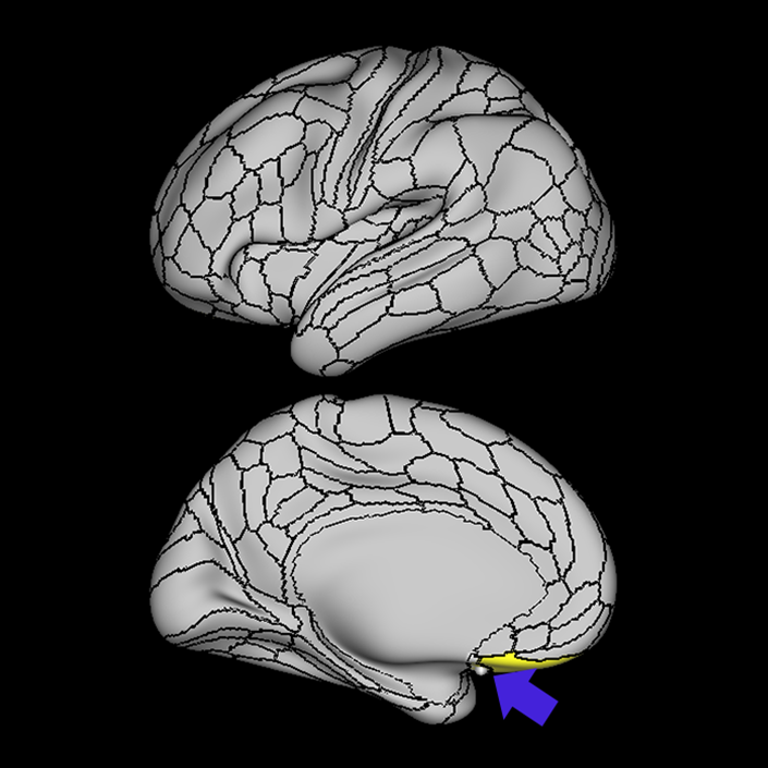
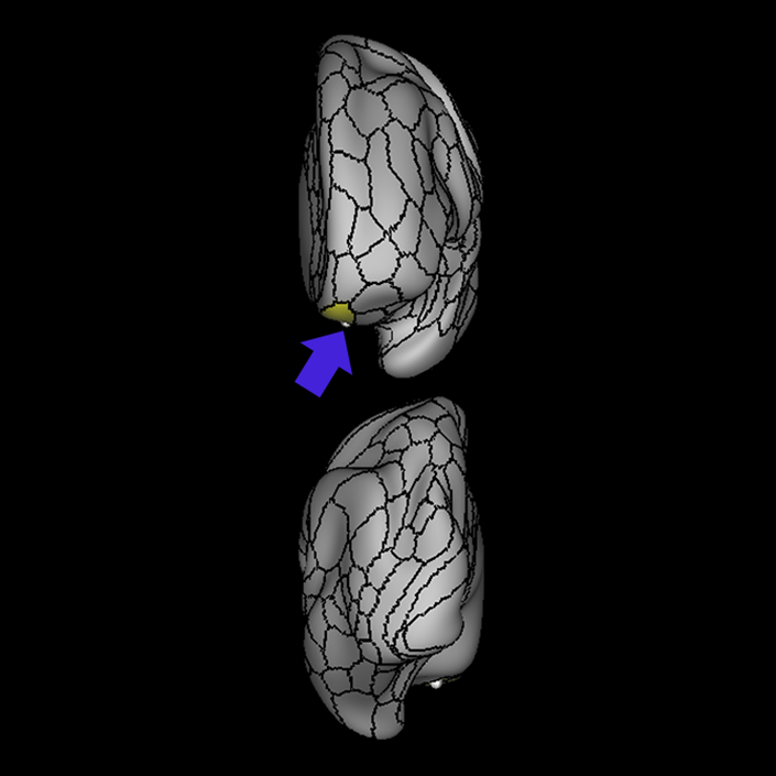
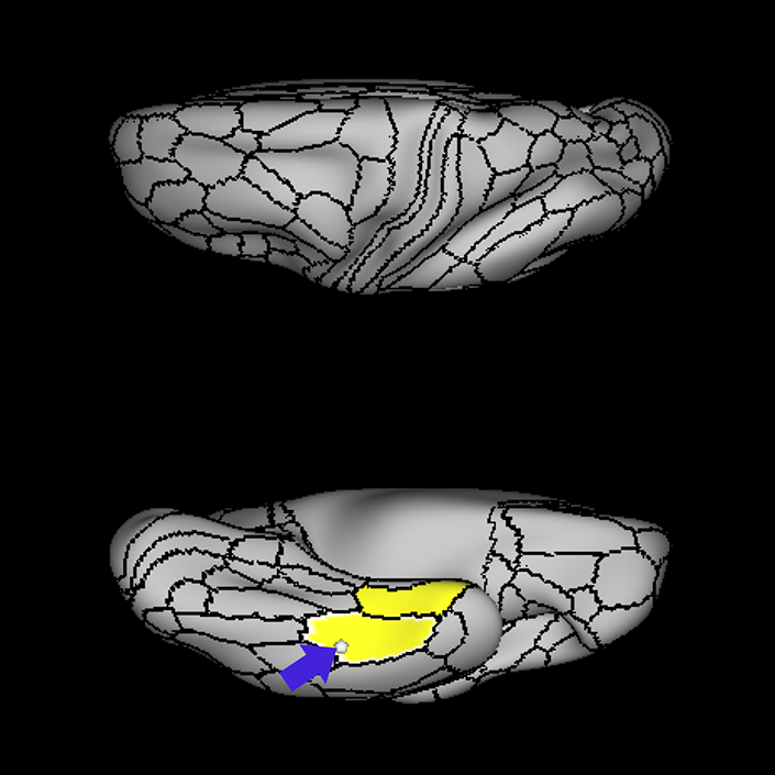
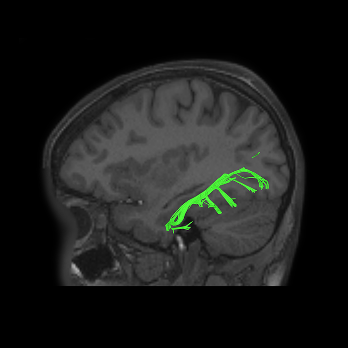
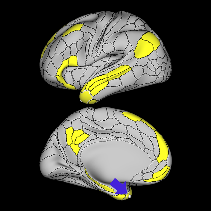
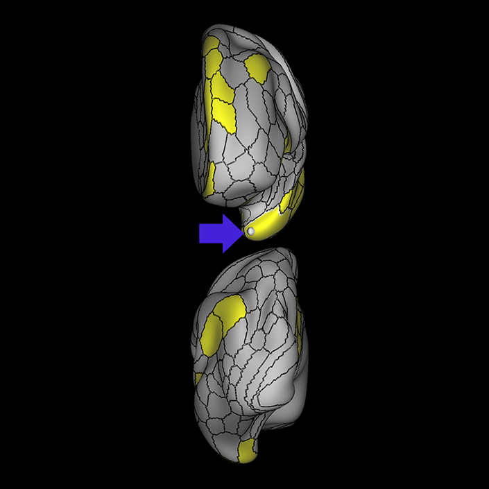
ᐅ SummaryArea OFC (orbitofrontal complex): part of the the orbitofrontal regions. Plays an integral role in the evaluation of rewards and punishment. Suggested to be involved in self- regulation, behavioral inhibition, and emotional control by predicting the moment-to-moment value of stimuli, actions, and choices based on internal states. ᐅ Where is it?Area OFC (Orbitofrontal complex) is located in the gyrus rectus, the medial orbitofrontal cortices, and the intervening sulcus. ᐅ What are its borders?Area OFC borders area 10pp anteriorly, areas 11l and 13l laterally, and area 10v on its superior medial bank. It abuts the pOFC posteriorly. ᐅ What are its functional connections?Area OFC demonstrates functional connectivity to areas pOFC and 13L. ᐅ What are its white matter connections?Area OFC is structurally connected to the contralateral hemisphere, the uncinate fasciculus and inferior fronto-occipital fasciculus. Contralateral connections course through the genu of the corpus callosum to end at 10d, 10r and 10v. Uncinate fasciculus fibers project through the limen insulae to temporal pole parcellation TGd. IFOF connections project posteriorly through the extreme/external capsule to occipital lobe area V1. ᐅ What is known about its function?The orbitofrontal cortex (Area OFC) plays an integral role in the evaluation of rewards and punishment. Additionally, the OFC is suggested to be involved in self-regulation, behavioral inhibition, and emotional control by predicting the moment-to-moment value of stimuli, actions, and choices based on internal states. |
|
A: lateral-medial
B: anterior-posterior
C: superior-inferior
DTI image |
ᐅ SummaryArea PeEc (perirhinal ectorhinal cortex): part of the temporal lobe regions. The perirhinal cortex: contributes to declarative memories transmitted between cortical areas and the hippocampus; adds semantic knowledge to aid in item identification; integrates item information with spatio-temporal information; transmits this data to the hippocampus via the EC. ᐅ Where is it?Area PeEC (perirhinal ectorhinal cortex) is found on the anterior portions and inferior surface of the uncus extending to the collateral sulcus. ᐅ What are its borders?Area PeEC borders areas TGv and TGd anteriorly and TF laterally. Its posterior borders are PHA2 and PHA3. ᐅ What are its functional connections?Area PeEC demonstrates functional connectivity to area IFSa the frontal lobe, areas EC, TF, PHA2, and PHA3 in the temporal lobe, area IP0 in the parietal lobe, and area PH in the occipital lobe. ᐅ What are its white matter connections?Area PeEC is structurally connected to the ILF. ILF projections travel through the inferior temporal lobe to end at PH, TPOJ3 and MT. In some individuals there are fibers that parallel the ILF to terminate at the medial occipital lobe at V1 ᐅ What is known about its function?The perirhinal cortex contributes to declarative memories transmitted between cortical areas and the hippocampus. The perirhinal cortex region adds semantic knowledge to aid in item identification. In addition, the perirhinal cortex integrates item information with spatio-temporal information and transmits this data to the hippocampus via the entorhinal cortex. The HCP authors were unable to reliably separate the perirhinal cortex and ectorhinal cortex, and therefore combined them as a single region. Area PeEC can be distinguished from neighboring regions based on increased activation in working memory primary task contrasts and increased activation in selective recognition of faces. Due to its particular affinity for facial recognition tasks compared to its neighbors, the HCP authors hypothesize that the region may be the site of the anterior temporal face patch |
|
A: lateral-medial
B: anterior-posterior
C: superior-inferior
DTI image |


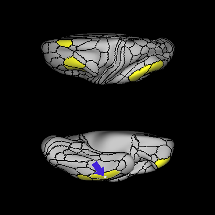
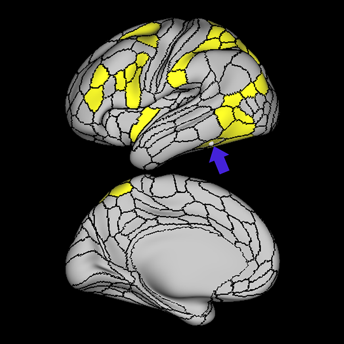
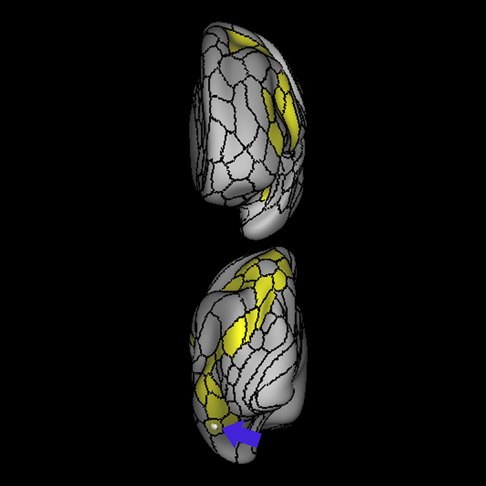
ᐅ SummaryArea pOFC (posterior orbitofrontal complex): part of the the orbitofrontal regions. Thought to play a role in behavior and decision- making by evaluating sensory reinforcers, such as taste and odor. ᐅ Where is it?Area pOFC (posterior orbitofrontal complex) is located in the posterior gyrus rectus and the medial posterior most portions of the orbitofrontal cortices. ᐅ What are its borders?Area pOFC borders OFC anteriorly, areas 47s and Pir laterally and area 25 superomedially. Its posterior border is with the anterior perforated substance. ᐅ What are its functional connections?Area pOFC demonstrates functional connectivity to the OFC. ᐅ What are its white matter connections?Area pOFC is connected to local parcellations. Short association bundles connect to the temporal pole through the insula to end at area TGd. There are anterior projections to OFC, 13l and PeEc. ᐅ What is known about its function?This posterior parcellation of the orbitofrontal cortex is thought to play a role in behavior and decision-making by evaluating sensory reinforcers, such as taste and odor. |
|
A: lateral-medial
B: anterior-posterior
C: superior-inferior
DTI image |
ᐅ SummaryArea PreS: (presubiculum) part of the medial temporal areas. Region of the hippocampus that primate studies suggest is involved in the processing of spatial information. Demonstrates less activity during tasks related to working memory, language processing, and theory of mind. Relative to PHA1, PreS shows greater activity during motor tasks. ᐅ Where is it?Area PreS is found on the posterior superior surface of the parahippocampal gyrus. ᐅ What are its borders?Area PreS borders the hippocampus medially, and the entrorhinal cortex anteriorly. Its posterior border is made up of RSC (retrosplenial cortex) and the ProS (prostriate region) (which are discussed in other sections. PHA1 is its inferior border ᐅ What are its functional connections?Area PreS demonstrates functional connectivity areas 8AD and i6-8 in the frontal lobe, areas PHA1, PHA2, and the hippocampus in the temporal lobe, areas RSC, Pros, d23ab, v23ab, 31a, 31pv, 7m, 7pm, POS1, POS2, IP1, and PGs in the parietal lobe, and area V1 in the occipital lobe ᐅ What are its white matter connections?Area PreS is structurally connected to the cingulum, precuneus and occipital lobe. Cingulum projections run superior to the corpus callosum to end at anterior cingulate cortex and frontal lobe parcellations a24, 9m, 10d and p32. There arePreS fibers that project posteriorly to end at occipital and precuneus areas V1, V2, V6, POS1, POS2 and 7m. Local short association fibers are connected to EC and PeEc. ᐅ What is known about its function?The presubiculum lies medial to the subiculum - a region of the hippocampus which primate studies suggest is involved in the processing of spatial information. Area PreS contains more myelin than the hippocampal cortex, and relative to PHA1 inferiorly, contains more myelin, is thinner, and demonstrates less activity during tasks related to working memory, language processing, and theory of mind. Relative to PHA1, PreS shows greater activity during motor tasks. |
|
A: lateral-medial
B: anterior-posterior
C: superior-inferior
DTI image |
ᐅ SummaryArea TE2a (temporal area 2 anterior): part of the temporal lobe regions. Appears primarily related to visual pathways. TE2a has a similar functional profile to TE1m, which borders the region superiorly, including activation in the visual working memory secondary contrast and deactivation in language tasks. Relative to TE1m, however, TE2a demonstrates less activation in visual working memory tasks and less deactivation during language tasks. ᐅ Where is it?Area TE2a (TE 2 anterior) is found on the anterior portion of the ITG, the anterior half of the inferior sulcus, and the lateral bank of the occipitotemporal sulcus. ᐅ What are its borders?Area TE2a borders TF on its basal-medial edge and TE1a and TE1m on its superior surface. It borders TGd and TGv anterior and its posterior end is wedged between TE1p and TE2p ᐅ What are its functional connections?Area TE2a demonstrates functional connectivity to areas 8AV, 8BL, 8C, and a47r in the frontal lobe, areas STSvp, TE1m, and TE1p in the temporal lobe, and areas PGs, PGi, and PFm in the parietal lobe. ᐅ What are its white matter connections?Area TE2a is structurally connected to the arcuate/SLF and ILF. ILF projections are inconsistent across individuals. Arcuate/SLF tracts wrap around the Sylvian fissure projecting toward the frontal lobe and turn medially to terminate at 6r, 6v, 8C, p9-46v, IFJa, IFJp and IFSp. There are posterior projections from the arcuate/SLF that terminate at the inferior parietal lobule at PF and PFm. Local short association fibers connect to TE1p and TGd Figure. ᐅ What is known about its function?The function of area TE2a appears primarily related to visual pathways. TE2a has a similar functional profile to TE1m, which borders the region superiorly, including activation in the visual working memory secondary contrast and deactivation in language tasks. Relative to TE1m, however, TE2a demonstrates less activation in visual working memory tasks and less deactivation during language tasks. |
|
A: lateral-medial
B: anterior-posterior
C: superior-inferior
DTI image |
ᐅ SummaryArea TE2p (temporal area 2 posterior): part of the temporal lobe regions. Appears primarily related to visual pathways. Relative to TE2a, TE2p is more active in theory of mind tasks and motor tasks. Notably, compared to the other TE1 and TE2 regions which are deactivated in the TOOL-AVG contrast, TE2p is activated unilaterally on the left in TOOL-AVG, demonstrating a possible role in object recognition. ᐅ Where is it?Area TE2p (TE 2 posterior) is found in the posterior part of the occipital temporal sulcus and the middle posterolateral portion of the fusiform gyrus. ᐅ What are its borders?Area TE2p borders PH posteriorly, and FFC medially. Its anterior border is TF and its lateral border is made up of TE2a, and TE1p. ᐅ What are its functional connections?Area TE2p demonstrates functional connectivity to areas FEF, PEF, IFSa, IFJa, IFJp, p9-46v, 6a, and 6r in the frontal lobe, area PoI2 in the insula, area PHT in the temporal lobe, areas PGp, AIP, MIP, LIPv, LIPd, IPS1, IP0, PFop, 7PC, 7PL, 7AL in the parietal lobe, and areas PH, FFC, FST, TPOJ2, and TPOJ3 in the occipital lobe. ᐅ What are its white matter connections?Area TE2p is structurally connected to the arcuate/SLF and local parcellations. White matter tracts of this parcellation are variable across individuals. Arcuate/SLF tracts wrap around the Sylvian fissure projecting toward the frontal lobe. The termination of the arcuate/SLF is unable to be delineated, as the tracts cannot be traced to specific parcellations. Local short association fibers connect to FFC, PH, TE2p, FFC, TE1m, TF and TE2a. White matter tracts in the right hemisphere of TE2p have consistent occipital connections. ᐅ What is known about its function?The function of area TE2p appears primarily related to visual pathways. Relative to TE2a, TE2p is more active in theory of mind tasks and motor tasks. Notably, compared to the other TE1 and TE2 regions which are deactivated in the TOOL-AVG contrast, TE2p is activated unilaterally on the left in TOOL-AVG, demonstrating a possible role in object recognition. |
|
A: lateral-medial
B: anterior-posterior
C: superior-inferior
DTI image |
ᐅ SummaryArea TF (temporal area f): part of the temporal lobe regions. Single neuron primate studies suggest that area TF may be involved in the maintenance of working memory in conjunction with inferior temporal and prefrontal regions. Primate studies also show that removal of the posterior parahippocampal region (containing areas TF and TH) produces consistent deficits in spatial tasks involving object¨Cplace association. In humans, TF is located in the anterior fusiform gyrus, which is important for visual perception such as in facial recognition, object recognition, and reading. Relative to TE2a, area TF is more activated in motor tasks, language recognition tasks, and theory of mind tasks. Relative to TE2p posteriorly, area TF demonstrates greater activity in language recognition tasks and facial recognition tasks. ᐅ Where is it?Area TF is found on the anterior part of the fusiform gyrus and the occipitotemporal sulcus. It occupies part of the lateral bank of the collateral sulcus. ᐅ What are its borders?Area TF borders TGv anteriorly, TE2a and TE2p laterally, PeEC medially, and FFC, VVC, PHA2 and PHA3 posteriorly. ᐅ What are its functional connections?Area TF demonstrates functional connectivity to PeEC and TE2p ᐅ What are its white matter connections?Area TF is structurally connected to the arcuate/SLF and ILF. The arcuate/SLF tracts wrap around the Sylvian fissure projecting toward the frontal lobe and turn medially to terminate at IFSa and 46. ILF terminations course through the inferior temporal lobe to terminate at V2, V3, V4, V3A and V3b. Local short association bundles connect to PeEc, VVC, TE2p and Te2a. ᐅ What is known about its function?Single neuron primate studies suggest that area TF may be involved in the maintenance of working memory in conjunction with inferior temporal and prefrontal regions. Primate studies also show that removal of the posterior parahippocampal region (containing areas TF and TH) produces consistent deficits in spatial tasks involving object-place association. In humans, TF is located in the anterior fusiform gyrus which is a hominid specific structure. The fusiform gyrus is important for visual perception such as in facial recognition, object recognition, and reading. The TE2 areas and TF comprise the ITG. Relative to TE2a which borders it laterally and superiorly, area TF is more activated in motor tasks, language recognition tasks, and theory of mind tasks. Relative to TE2p posteriorly, area TF demonstrates greater activity in language recognition tasks and facial recognition tasks. |
|
A: lateral-medial
B: anterior-posterior
C: superior-inferior
DTI image |
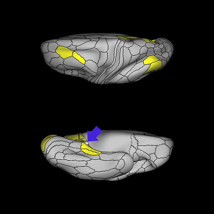
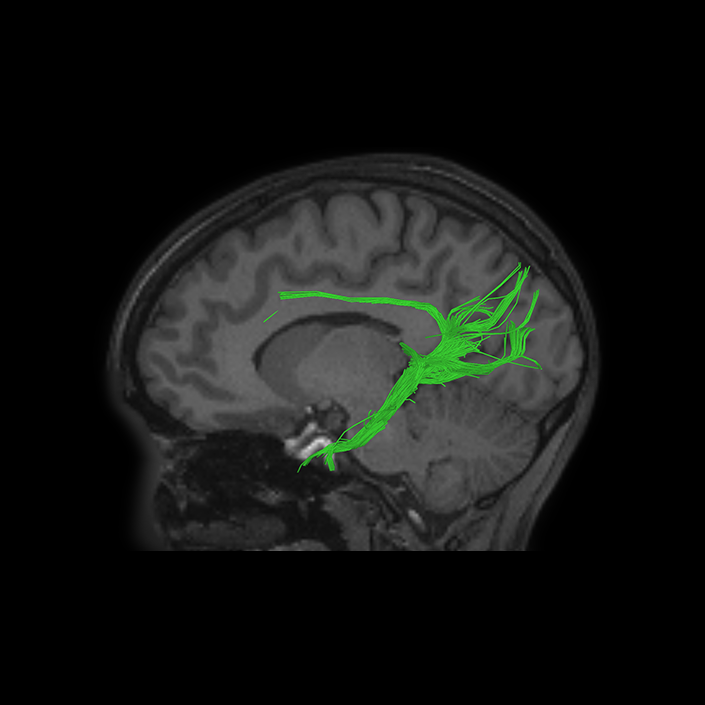
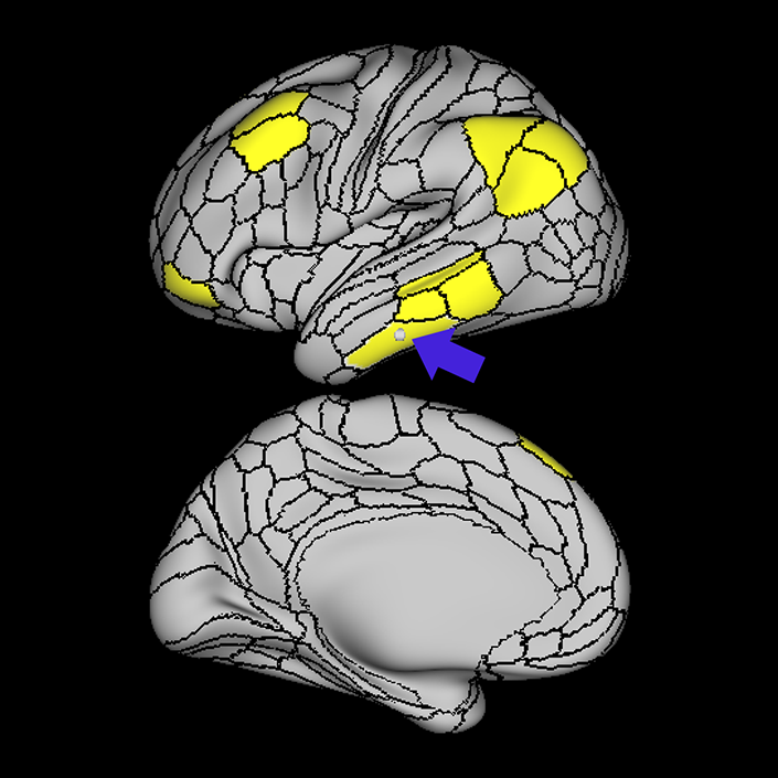
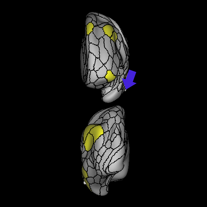
ᐅ SummaryArea TGd (temporal area g dorsal): part of the temporal lobe regions. Areas TGd and TGv make up the temporal polar cortex, a paralimbic region important for social and emotional processing, auditory and visual aspects of facial recognition, emotional processing of auditory, olfactory and visual stimuli, and theory of mind. TGd, like TGv, is activated in the language-related task contrasts suggesting a role in ventral stream language processing along with its neighbors STGa, STSda, STSva, and TE1a. Compared to TGv inferiorly, TGd is deactivated vs activated in motor tasks in response to a visual cue and relational primary contrasts (ie, distinguishing objects based on feature dimensions). ᐅ Where is it?Area TGd (TG dorsal) is found on the superior part of the temporopolar region. It is roughly anterior to the STG and MTG and wraps over the superior surface of the temporal planum polare just anterior to the limen insula. ᐅ What are its borders?Area TGd borders STGa, STSda, and TE1a posteriorly, TE2a and TGv inferiorly, Pir and PI on its anteromedial edge, and PeEC on its posterior mesial border. ᐅ What are its functional connections?Area TGd demonstrates functional connectivity to 8AV, 8BL, 9a, 9p, 9m, 44, 45, 47s, 47L, 10v, 10r, and SFL in the frontal lobe, areas STSva, STSvp, STSda, STSdp, TGv, STGa, PeEc, the hippocampus, and TE1a in the temporal lobe, and areas PGs, PGi, 7m, d23ab, 31pv, and 31pd in the parietal lobe. ᐅ What are its white matter connections?Area TGd is structurally connected to the uncinate fasciculus and ILF. Many individuals also have connections through the extreme/external capsule toward the parieto-occipital sulcus and occipital lobe. The uncinate fasciculus wraps medially around the fibers travelling toward the occipital lobe, these fibers course through the extreme/external capsule through the posterior temporal lobe to terminate at DVT, V1, V3, V2, V6 and 7PL. The uncinate fasciculus projects to the frontal lobe through the insula to terminate at FOP4, FOP5, 44 and 45. ILF fibers course through the inferior temporal lobe to terminate at V1 and V2. ᐅ What is known about its function?Areas TGd and TGv make up the temporal polar cortex, a paralimbic region important for social and emotional processing, auditory and visual aspects of facial recognition, emotional processing of auditory, olfactory and visual stimuli, and theory of mind. TGd, like TGv, is activated in the language-related task contrasts suggesting a role in ventral stream language processing along with its neighbors STGa, STSda, STSva, and TE1a. Compared to TGv inferiorly, TGd is deactivated vs activated in motor tasks in response to visual cue and relational primary contrasts (i.e. distinguishing objects based on feature dimensions). |
|
A: lateral-medial
B: anterior-posterior
C: superior-inferior
DTI image |
ᐅ SummaryArea TGv (temporal area g ventral): part of the temporal lobe regions. Areas TGv and TGd make up the temporal polar cortex described in the previous section. Area TGv, like TGd, is activated in language-related task contrasts suggesting a role in ventral stream language processing. Compared to TGd superiorly, TGv is activated vs deactivated in motor tasks in response to a visual cue and relational primary contrasts (ie, distinguishing objects based on feature dimensions). ᐅ Where is it?Area TGv (TG ventral) is found on the inferior temporal polar region just anterior to the ITG and fusiform gyrus. ᐅ What are its borders?Area TGv borders TE2a posteriorly on its lateral surface, TF posteriorly on its basal surface, and TGd superiorly. PeEc makes up its medial basal surface. ᐅ What are its functional connections?Area TGv demonstrates functional connectivity to areas 45 and TGd. ᐅ What are its white matter connections?Area TGv is structurally connected to the ILF. ILF projections travel through the inferior temporal lobe to terminate at V1, V2, V3 and V4 ᐅ What is known about its function?Areas TGv and TGd make up the temporal polar cortex described in the previous section. Area TGv, like TGd, is activated in language-related task contrasts contrasts suggesting a role in ventral stream language processing. Compared to TGd superiorly, TGv is activated vs deactivated in motor tasks in response to a visual cue and relational primary contrasts (i.e. distinguishing objects based on feature dimensions) |
|
A: lateral-medial
B: anterior-posterior
C: superior-inferior
DTI image |